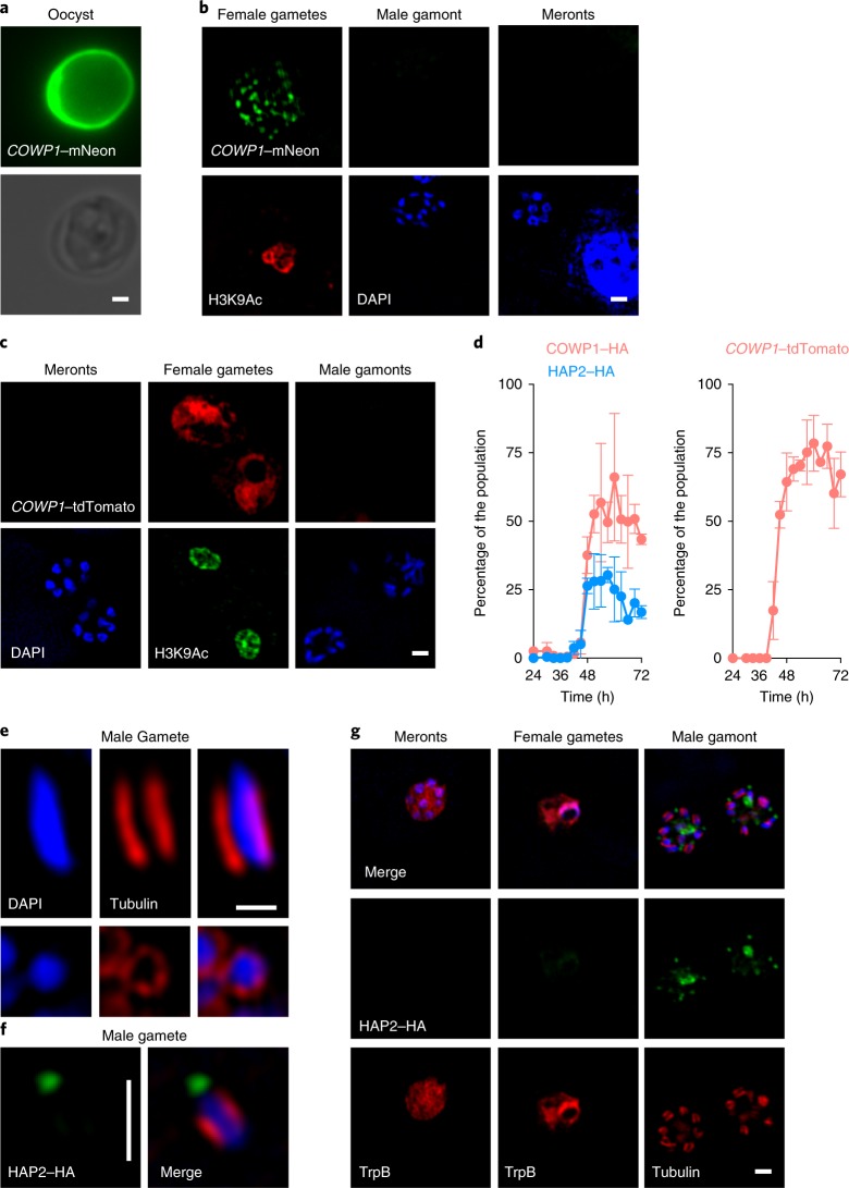
Fig. 2. Exclusive molecular markers for the sexual stages of C. parvum.
a–d, C. parvum were engineered to express COWP1–mNeon (a,b) and COWP1–HA (Supplementary Fig. 3) from the native locus or COWP1-promoter-driven tdTomato from the ectopic TK locus (c). Note the mNeon labelling of the wall in oocysts purified from infected mice and punctate labelling in female gametes observed in infected HCT-8 cells. Labelling becomes apparent after 42 h of culture and is never observed in asexual meronts or male gametes (b,d). The COWP1 promoter alone is sufficient to confer female-specific expression to a reporter protein (c,d). Anti-H3K9Ac antibodies were used to label the nuclei of females because they stain poorly with 4,6-diamidino-2-phenylindole (DAPI). For the time intervals in d, cultures were infected with the indicated transgenic strains and triplicate coverslips were fixed and processed for immunofluorescence assays. Parasite stages were scored for HA staining, the mean ± s.d. percentage of HA-positive stages among all of the parasites is shown for three independent biological replicates. e, Male gametes show a characteristic array of microtubules around the nucleus after staining with anti-tubulin antibodies. f,g, When parasites were engineered to express HAP2–HA from the native locus, antibody staining revealed exclusive labelling of free gametes (f) and male gamonts (g). HAP2 labels a single pole per mature gamete. This staining becomes apparent after 42 h of culture (d, blue). All of the microscopy experiments shown in this figure were performed independently three times. Scale bars in a–c,f,g, 1 µm; scale bar in e, 0.5 µm.

