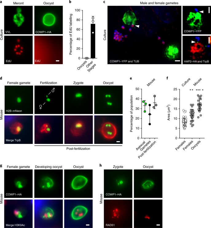
Fig. 5. Cryptosporidium males locate females in culture, but fertilization and meiosis only occur in vivo.
a,b, HCT-8 cells were infected with COWP1–HA C. parvum and after 36 h of infection, the nucleotide analogue EdU was added to the medium. Then, 12 h later, cells were click labelled and counterstained with anti-HA antibodies or Vicia villosa lectin (VVL; a). Cells were scored for nuclear EdU labelling (b); 100 stages were quantified for three biological replicates, and the experiment was performed twice. Data are mean ± s.d. c, Representative images of encounters between male and female gametes in culture; gametes were identified using the indicated transgenes or antibodies, and attached males are highlighted by arrowheads. YFP, yellow fluorescent protein. d–h, Ifng−/− mice were infected with H2B–mNeon-expressing (d) or COWP1–HA-expressing (g,h) parasites, and intestines were sectioned and prepared for immunofluorescence assays and counterstained with anti-TrpB, anti-H3K9Ac or anti-RAD51 antibodies. Representative micrographs show progression of events following fertilization. Post-fertilization stages are abundant in vivo (e) and these stages were significantly larger than those found in vitro (f); each symbol represents a parasite, n = 25. For e, data are mean ± s.d. from three independent mice. For f, data are mean ± s.d.; the statistical analysis was performed using a two-sided Student’s t-test comparing cultured females with in vivo females (**P = 0.0023) or with in vivo oocysts (****P = 0.0001). All of the microscopy experiments shown in d–h were performed twice with similar results. Scale bars, 1 µm.

