Figure 3.
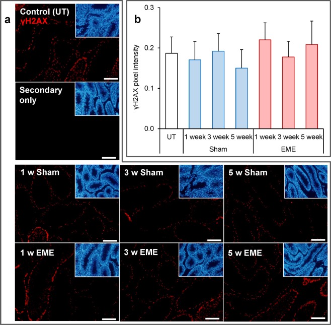
RF-EME exposure does not induce γH2AX expression in the testis. Testis sections from untreated control animals (UT), as well as those of the sham and RF-EME exposure groups, were probed with anti-phospho-γH2AX antibodies (red) to detect DNA double strand breaks. (a) Representative images are depicted, with scale bar equating to 400 µm. A secondary antibody only control is also included. Corresponding DAPI (blue) stained images, illustrating tubule morphology are included as insets included in the upper right corner of each panel. (b) Analysis of pixel intensity was performed on the germ cell population within the seminiferous tubules in order to quantify γH2AX expression levels across treatments. Graphical data are presented as mean + SEM (n = 3 mice/treatment group, with 8–25 tubules being analyzed for each testis).

