
Figure 6.
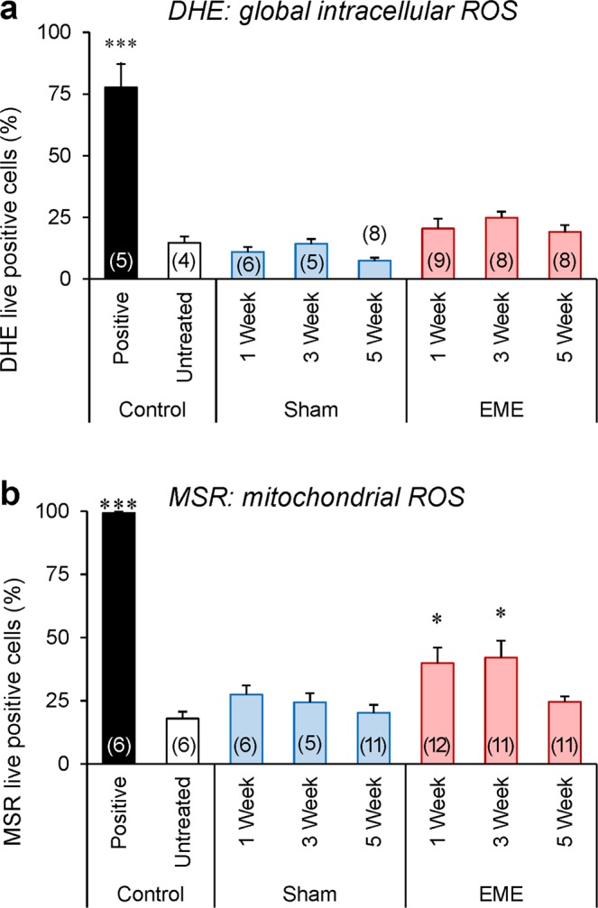
Exposure to RF-EME stimulates the generation of mitochondrial reactive oxygen species. Spermatozoa were isolated from the cauda epididymis of untreated control animals, as well as those of the sham and RF-EME exposure groups. These cells were pre-loaded with fluorescent probes and then analyzed using flow cytometry to assess their generation of reactive oxygen species (ROS). (a) Global levels of ROS generated in the sperm cell was assessed with the dihydroethidium (DHE) probe. (b) Alternatively, mitochondrial ROS generation was investigated with the MitoSOX Red (MSR) probe. In both instances, a minimum of 10,000 spermatozoa were assessed from 5–12 of animals and data are presented as mean + SEM. The number of biological replicates used is denoted in each bar. *P < 0.05.