Figure 7.
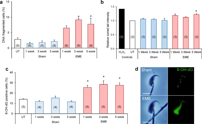
RF-EME exposure induces oxidative DNA damage in spermatozoa. Spermatozoa were isolated from the cauda epididymis of untreated control animals (UT), as well as those of the sham and RF-EME exposure groups. These cells were assessed for DNA fragmentation using (a) the halo assay showing the percentage of cells fragmented (n = 5–8 mice/treatment group, each with 100 sperm assessed for each replicate) and then (b) quantified by the alkaline comet assay, expressed as percentage tail intensity and normalized to control data for each run (n = 3 mice/treatment, each with 50–70 sperm cells assessed). (c) To extend this DNA integrity analysis, sperm were evaluated for oxidative DNA adducts via labelling with anti-8-hydroxy-2-deoxyguanosine (8-OH-dG) antibodies (n = 3–5 mice/treatment). (d) Representative images of spermatozoa stained with the 8-OH-dG antibody from the 5 week sham and RF-EME exposed populations are included. The number of biological replicates used is denoted in each bar. Data are presented as mean + SEM. *P < 0.05, **P < 0.01.

