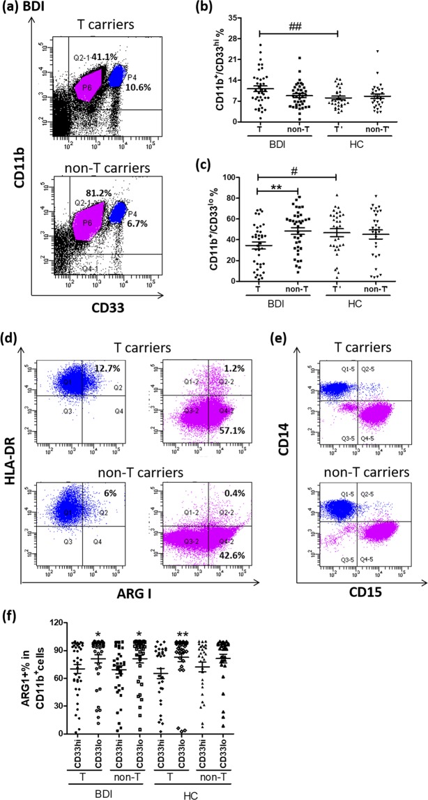
Figure 1.
Characterization of CD11b+ cells with flow cytometry. PBMCs were stained with antibodies against CD11b, CD33, and HLA-DR on ice for 30 min. After fixation and permeabilization, cells were incubated with an antibody against ARG1 on ice for another 30 min. (a) Stained cells were analyzed using a flow cytometer, which revealed two distinct populations of CD11b+/CD33hi (P4 gate) and CD11b+/CD33lo (P6 gate) cells from BDI patients. The percentages of (b) CD11b+/CD33hi and (c) CD11b+/CD33lo cells between BDI patients and healthy controls (HC) were calculated and adjusted for gender and age using general linear regression (#p < 0.05; ##p < 0.01). Among BDI patients, only the percentage of (c) CD11b+/CD33lo cells differed significantly between rs17026688 T and non-T carriers, as assessed with the Mann-Whitney test (**p < 0.01). These two groups of CD11b+ cells were further gated to examine the expression of HLA-DR and ARG1 (d) or of CD14 and CD15 (e). (f) The percentage of ARG1+ cells was compared between CD11b+/CD33hi and CD11b+/CD33lo populations in rs17026688 T and non-T carriers among BDI patients and HC using the Mann-Whitney test (*p < 0.05; **p < 0.01). Horizontal lines denote the mean ± SEM.

