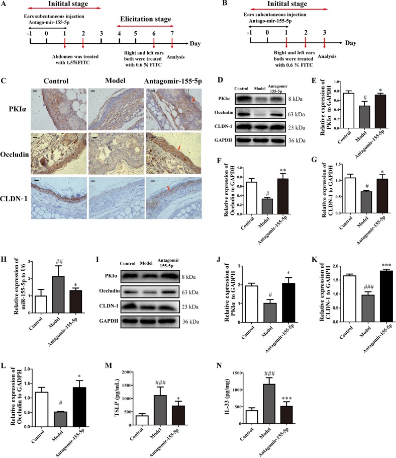
Fig. 6. Silencing miR-155-5p restored PKIα and TJ protein expression and reduced TSLP and IL-33 production in different stages of the AD model.
a, b Overview of the protocol used to establish the elicitation and initial stages of the FITC-induced AD mouse model. c, d PKIα, CLDN-1, and occludin expression was analyzed with immunohistochemistry (n = 5; magnification: ×630; scale bar: 50 μm) and with western blotting in the elicitation stage of the AD model. e–g PKIα, CLDN1, and occludin expression was quantified relative to GAPDH expression with a ChemiScope analysis. h, i Expression of miR-155-5p was detected with PCR and that of PKIα, CLDN-1, and occludin was analyzed with western blotting in the initial stage of the AD model. j–l PKIα, CLDN-1, and occludin expression was quantified relative to GAPDH expression with a ChemiScope analysis (n = 4). m, n TSLP and IL-33 production in ear homogenates in the initial stage of the AD model was measured with ELISAs (mean ± SD; n = 6; #P < 0.05, ##P < 0.01, ###P < 0.01 versus control, *P < 0.05, **P < 0.01, ***P < 0.001 versus model).

