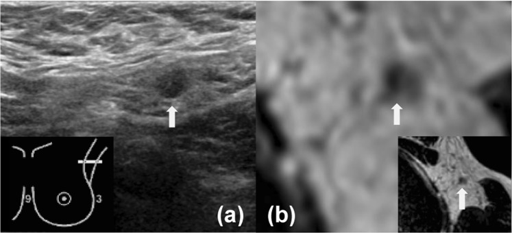
Figure 1.
Images of the axilla of a 52-year-old female patient with a 34 mm large invasive ductal carcinoma in her left breast, which was treated with mastectomy and ALND. For both US and MRI (reader 1 and 2) N1 axillary lymph node disease was reported. The white arrow indicates the suspicious lymph node on US and MRI. Histopathology of the ALND reported pN2–3 (largest diameter, 14 mm). (a) Axillary US (b) Transversal unenhanced T2-weighted breast MRI.

