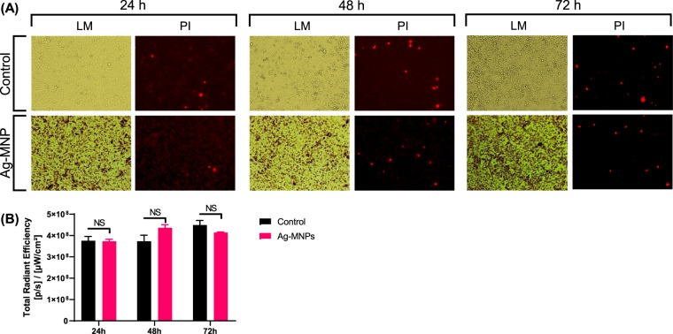
Figure 5.
In vitro cytotoxicity assessment of Ag-MNP. Hep-2 cells were cultured for 24, 48 and 72 h in EMEM-Glutamine + 10% FBS medium containing 0 μg/ml (Control) or 100 μg/ml of Ag-MNP. Micrographs in (A) indicate Light Microscopy (LM) and fluorescence imaging of Propidium Iodide (PI; ʎEx = 535 nm and ʎEm = 617 nm) that stained dead cells (Red color. The PI signal intensities were quantified in (B) using the In Vivo Imaging System (IVIS, Perking Elmer).

