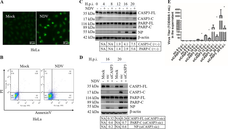
Fig. 1. NDV infection induces apoptosis in HeLa cells.
a Detection of NDV-induced apoptosis by TUNEL assay. HeLa cells were infected with NDV Herts/33 strain (MOI = 1) or mock-infected, and subjected to TUNEL assay at 20 h.p.i. The images of TUNEL positive cells were captured by a fluorescence microscope (×200). b Detection of NDV-induced apoptosis by Annexin V/PI staining and flow cytometry. HeLa cells were infected with NDV or were mock-infected, stained with annexin V and PI, and analyzed with flow cytometry at 20 h.p.i. c Detection of NDV-induced apoptosis in HeLa cells by western blot analysis. HeLa cells were infected with NDV or mock-infected, and harvested at 0, 4, 8, 12, 16, and 20 h.p.i. The cleavage of caspase-3 (CASP3) and PARP, the expression of NDV NP were determined by western blot analysis. The intensities of indicated protein bands were determined by image J, normalized to β-actin, and shown as fold change of NDV (+:−). d, e Knockdown of CASP3 by siRNA reduces apoptosis and virus release. HeLa cells were transfected with siCASP3 or sic for 36 h, followed by NDV infection. Mock infection without siRNA transfection was included as control. The cells lysates were analyzed with western blot with indicated antibodies (d). The intensities of indicated protein bands were determined, normalized to β-actin, and shown as fold change of siCASP3:sic. In parallel, the culture medium were collected at indicated time points and subjected to TCID50 assay (e). TUNEL assay, flow cytometry, and western blot are representative of three independent experiments. Virus titer represents means ± SD of three independent determinations. *p < 0.05.

