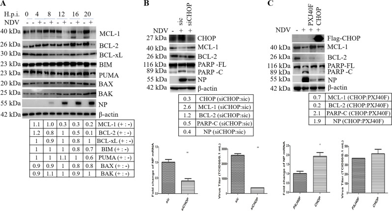
Fig. 4. CHOP promotes apoptosis by downregulation of anti-apoptotic protein BCL-2 and MCL-1 during NDV infection.
a Downregulation of BCL-2 and MCL-1 by NDV infection. HeLa cells were infected with NDV and harvested at indicated time points. Western blot analysis was performed to detect MCL-1, BCL-2, BCL-xL, BIM, PUMA, BAX, BAK, NP, and β-actin. The intensities of indicated protein bands were normalized to β-actin and shown as fold change of NDV (+:−). b, c CHOP regulates the levels of BCL-2/MCL-1, apoptosis, and virus proliferation. HeLa cells were transfected with sic or siCHOP for 36 h, or transfected with PXJ40F-CHOP or PXJ40F for 24 h, followed with NDV infection. Mock infection was set as control. Cells were harvested at 20 h.p.i. and analyzed with western blot (b, c, upper panels), or subjected to quantitative real-time RT-PCR to detect NP mRNA (b, c, low panels). In parallel, the cell culture medium was collected and subjected to TCID50 assay to measure the released progeny virus (b, c, low panels). The intensities of indicated protein bands were normalized to β-actin and shown as fold change of siCHOP:sic or CHOP:PXJ40F. Western blot results are representative of three independent experiments. NP mRNA level and virus titer represent means ± SD of three independent determinations. *p < 0.05, **p < 0.01.

