
Fig. 2. Cell cycle blockage and proapoptotic activity of BSB.
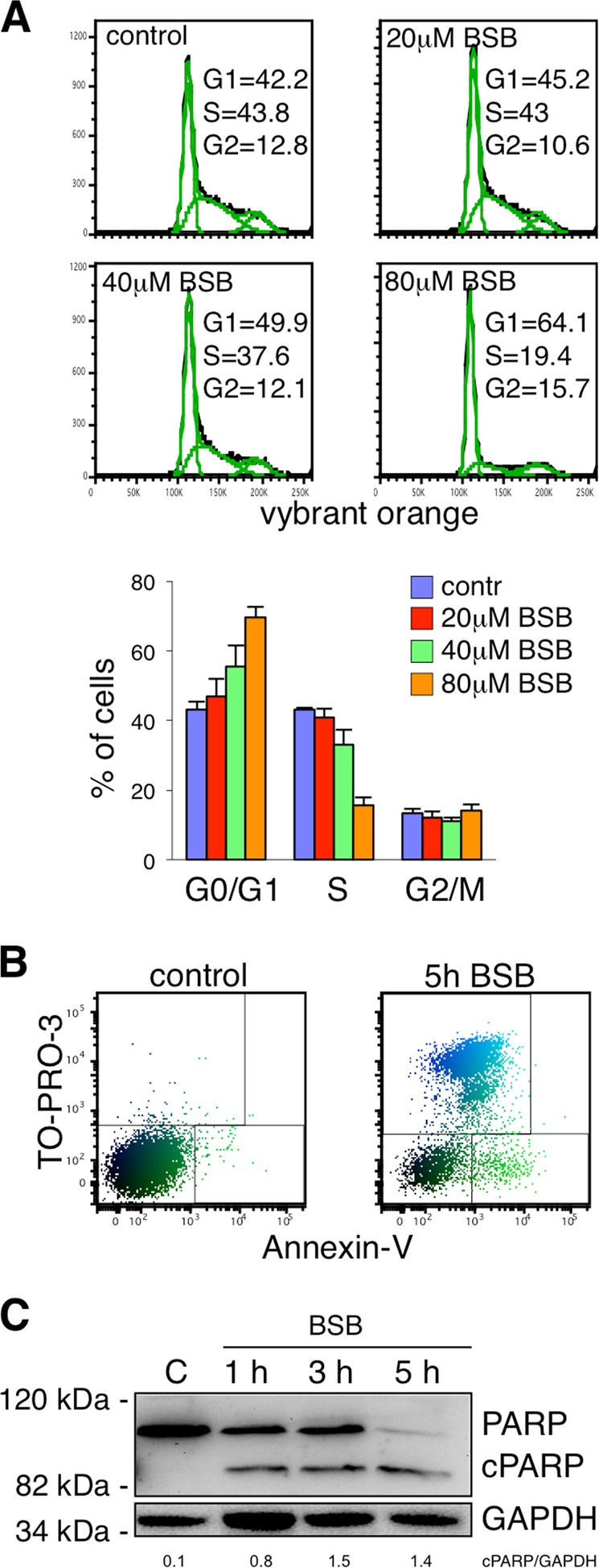
a Top: a representative flow cytometric experiment demonstrating cell cycle arrest by BSB. Jurkat cells treated with 20, 40, and 80 µM BSB for 24 h were incubated with 10 µM Vybrant DyeCycle orange stain for 15 min. Bottom: statistical analysis (mean and s.d.) of the cell cycle phases. b Cytometric PolyChromatic plot analysis of apoptosis after 5-h treatment with 80 µM BSB. Jurkat cells were stained with Annexin-V and TO-PRO-3 to distinguish between alive (Annexin-V−/TO-PRO-3−), early apoptotic (Annexin-V+/TO-PRO-3−), and late apoptotic/necrotic cells (Annexin-V+/−/TO-PRO-3+). c Western blot showing the appearance of the cleaved form of PARP (cPARP) following 1, 3 and 5 h treatment with 80 μM BSB in Jurkat cells. Each experiment is representative of five.