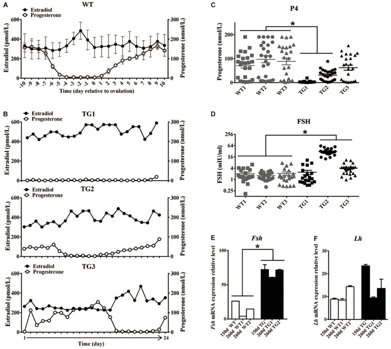
FIGURE 2.
Transgenic gilts presented disordered estrous cycle and reproductive hormones. Plasma E2 and P4 concentration of 365-day old WT and TG gilts were measured at a 24-h interval for 24 days. (A) During the estrous cycle, representative WT gilts showed typical serum E2 concentration peak before ovulation, accompanied with marked decreased P4 concentration (n = 3), data are presented as mean ± standard deviation. (B) Irregular plasma E2 concentration peaks was observed in three representative TG gilts in a continuous 24-day measurement. (C) The average P4 concentration of two of the three TG gilts in a continuous 24-day measurement was significantly lower than that of WT gilts (p < 0.05). Each point stands for a P4 concentration value. (D) Two of the three 365-day TG gilts showed higher serum FSH concentration. Serum FSH concentration was measured at a 24-h interval for 24 days. (E) Expression level of Fsh mRNA in the pituitary of both 150- and 260-day old TG gilts were two-fold higher than that in WT gilts (p < 0.05). (F) The average level of Lh mRNA level in pituitary was not significantly different in TG and WT gilts. ∗Stands for statistical significance (P < 0.05).

