FIGURE 3.
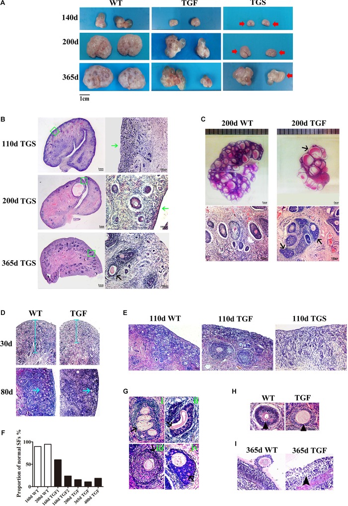
BMP15 knockdown-induced changes in ovarian and follicular development. (A) Representative photograms of ovaries collected from gilts of different ages showing reduced ovarian size and less visible follicles on the surface of TG ovaries as compared to ovaries from WT sibling. Bilateral TG ovaries were significantly different in size at ages of 200 and 365 days. Two ovarian phenotypes were found in TG ovaries, TGF ovaries had many visible large antral follicles on the ovarian surface. TGS ovaries contained none or less than three visible antral follicles (red arrows). (B) Histological observation of TGS ovaries showed that 110- and 200-day old TGS ovary presented major primary-like follicles sparsely scattered on the cortex (green arrows), while 365-day old TGS ovarian section was predominantly occupied by degraded secondary follicles (black arrow). (C) On the 200-day TGF ovarian section, decreased number of follicles and enlarged antral follicles (black arrow) was observed. In addition, the degradation of GCs in abnormally organized GC layer structure of secondary follicles was observed (black arrows). (D) In 30- and 80-day old TGF ovaries, drastically decreased number of early stage follicles led to thinner ovarian cortex (blue line and blue arrows). (E) Comparison of three ovarian phenotypes at the age of 110 days showed less number of early stage follicles in TGF ovarian cortex, and the minimum number of follicles in TGS ovaries. (F) Markedly reduced proportion of normal secondary follicles in the TGF ovaries. Secondary follicles in three sections of each ovary were counted. (G) Representative images of abnormal TGF secondary follicles (black arrows), including multiovular follicle with highly irregularly organized theca cell layers (i); follicle with oocyte-free structure, and abnormally thickened zona pellucida surrounded by highly degraded GCs (ii); follicle with abnormally thickened theca layers (iii); follicle with enlarged oocyte surrounded by highly irregularly organized GC layers with holes formed by degradation of GCs (iv). (H) TGF follicle showed larger oocyte in the early secondary follicle stage (black arrow head). (I) Smaller GCs were loosely organized in TGF antral follicles (black arrow head).

