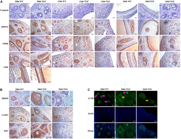
FIGURE 5.
Abnormal TGF follicles showed premature luteinization and impaired oocyte quality. (A) Immunostaining of ovarian sections indicated that the expression of BMP15 decreased remarkably in TGS abnormal follicles, but was only slightly reduced in TGF follicles, compared with WT follicles. FSHR shared a similar expression pattern with BMP15, and the expression pattern of LHR was different from that of BMP15. It expressed higher in TGF GCs of both preantral and antral follicles. Scale bar = 100 μm. (B) Follicular cell apoptosis, proliferation, and premature luteinization were evaluated by immunostaining with Caspase3, Ki67, and 3βHSD, respectively. Notably, higher expression level of 3βHSD was discovered in abnormal TGF follicles. However, the expression of Caspase3 and Ki67 in abnormal TGF follicles was not significantly different from that in WT follicles. Scale bar = 100 μm. (C) Immunofluorescence images demonstrates intensive expression of autophagy-related protein, LC3B, in oocytes of normal follicles of TGF and WT ovaries, but it was barely expressed in oocytes of abnormal follicles of TG (TGF and TGS) ovaries. Purple arrow, oocytes in normal follicles; Orange arrow, oocytes in abnormal follicles. Scale bar = 100 μm.

