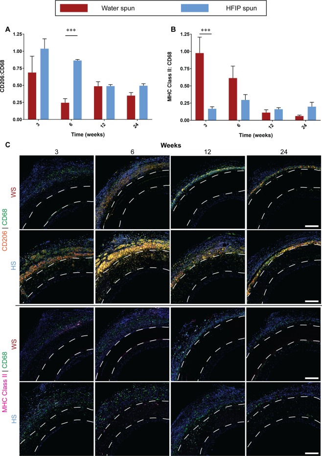
Figure 7.
Macrophage response to electrospun silk grafts. Data expressed as mean ± SEM, n = 4 animals/timepoint. Statistical analysis comparing these averages was by two-way ANOVA, using Šídák multiple comparison test, with *p < 0.05, **p < 0.01, ***p < 0.001. (A) Quantification of M2 polarisation represented as a ratio of CD206:CD68. (B) Quantification of M1 polarisation represented as a ratio of MHC Class II:CD68. (C) Representative images of cross sections at the proximal end of HS silk vascular grafts, white dotted lines indicate the graft wall. Upper two rows: CD206 stained in orange, CD68 stained in green and nuclei stained in blue. Lower two rows: MHC Class II stained in purple, CD68 stained in green and nuclei stained in blue. Scale bar = 200 μm.

