Figure 3.

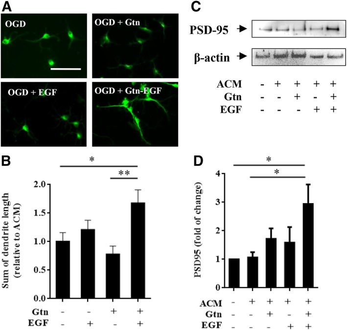
Epidermal growth factor (EGF)‐hydrogel treated astrocyte conditioned media (ACM) promoted dendritic regeneration and synaptogenesis in primary neurons. (A): Representative MAP‐2 staining (green, for dendrites) on primary neuron after subjected to 2 hours oxygen–glucose deprivation (OGD) with 22 hours neuron conditioned medium (control) or ACM from astrocytes treated with gelatin hydrogel (Gtn), EGF, or EGF‐hydrogel (Gtn‐EGF) under normoxia condition. Scale bar = 50 μm. (B): Sum of primary neuron dendritic length after subjected to 2 hours OGD followed by 22 hours of ACM collected from astrocytes treated with Gtn, EGF, or Gtn‐EGF under normoxia condition. Error bars represent SEM, n = 20. (C): Representative images of PSD‐95 (postsynaptic density markers) and β‐actin (housekeeping gene) expression of primary neuron after subjected to 2 hours OGD and followed by 22 hours of neuron conditioned medium (−ve ACM, Gtn, and EGF) or ACM treated with Gtn, EGF, or Gtn‐EGF under normoxia condition. (D): The densitometry measurement for PSD‐95 of primary neuron after subjected to 2 hours OGD and followed by 22 hours of neuron conditioned medium (−ve ACM, Gtn, and EGF) or ACM treated with Gtn, EGF, or Gtn‐EGF under normoxia condition. Data were normalized to primary neuron subjected to 2 hours OGD followed by 22 hours of neuron conditioned medium treated for 22 hours under normoxia condition. Error bars represent SEM, n = 5; *, p < .05; **, p < .01.
