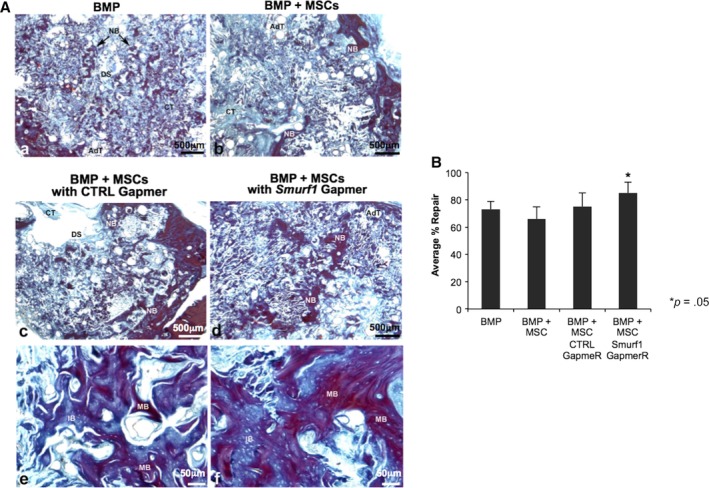
Figure 2.

Bone regeneration in a calvarial critical size defect. (A): Sixteen female ovariectomized Sprague–Dawley were divided in four experimental groups of four rats each (n = 4). (A–D): Semipanoramic representative images showing in the experimental groups BMP (A), BMP Cells (B), BMP Cells LNA‐ASO Ctrl, (C) and BMP Cells LNA‐ASO Smurf1 (D) the repair response where newly formed bone (NB) can be seen both on the margins and inside the defect. The images (E) and (F) show details at high magnification of the newly formed bone in the BMP Cells LNA‐ASO Ctrl (E) and BMP Cells LNA‐ASO Smurf1 (F) groups. Observe the greater homogeneity and continuity in the structure of the bone in the group BMP Cells LNA‐ASO Smurf1 as well as larger areas of mineralization stained red (mature bone). Abbreviations: AdT, adipose tissue; CT, connective tissue; DS, defect site; NB, newly formed bone; IB, inmature bone; MB, mature bone. (A–D) ×25, (E, F) ×200. (B) Histomorphometrical analysis. Comparison of the percentages of repair among the different experimental groups by means of a one‐way ANOVA with Tukey multiple comparison posttest. Bar graph depicts percentages of repair at different experimental time points. Bars represent means ± SD, *p < .05.
