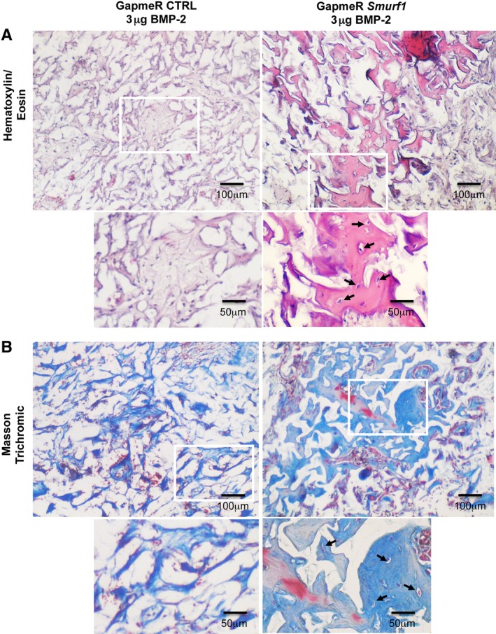
Figure 3.

Hematoxylin/eosin and Masson‐Goldner staining of subdermical implants in rats pictures show histological analysis of sections obtained from decalcified implants (n = 4). Hematoxylin/eosin (A) and Masson–Goldner staining (B) showing mineralized matrix in light pink and light blue respectively. Arrows indicate osteocytes‐like cells surrounded by lacunae and immersed in the mineralized matrix. Magnification: ×4 and ×8.
