Figure 3.

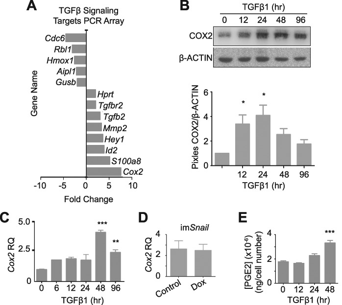
mOSE cells treated with TGFβ1 (10 ng/mL) increase Cox2 expression and PGE2 secretion. A–C: mOSE cells treated with TGFβ1 (4 days) have increased Cox2 expression as determined by a TGFβ1 Signaling Targets PCR array (A, N = 3, Student t-test), and validated by western blot (B, representative western blot, N = 3, One-way ANOVA with Dunnett’s post-test) and Q-PCR (C, N = 3, One-way ANOVA with Dunnett’s post-test). β-ACTIN was used as a loading control for the western blot. mOSE cells were treated with TGFβ1 up to 96 h. D: Cox2 expression is unaffected in imSnail mOSE cells treated with doxycycline (200 ng/mL, 48 h) as determined by Q-PCR (N = 3, Student t-test). E: mOSE cells treated with TGFβ1 have increased PGE2 production at 48 h after treatment, compared to untreated mOSE cells, determined by a PGE2-ELISA (N = 3, One-way ANOVA with Dunnett’s post-test). ** and *** indicate significant differences from the untreated control, P < 0.01 and P < 0.001 respectively. RQ = relative quantity. Experiments were performed using cells under passage number 25.
