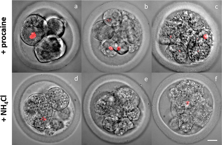
Figure 6.

Combined lightmicroscopic and DNA-fluorescence confocal micrographs of embryo developmental stages (DNA configuration in red) (A and D: 2–4 cell; B and E: 8-cell; C and F: 16-cell) after oocytes were incubated for 18 h in CM + 2.5 mM procaine and CM + 25 mM NH4Cl, and subsequently cultured in a DMEM/F12 plus 10% FBS based medium. Oocytes that cleaved in conditions containing 2.5 mM procaine and 25 mM NH4Cl never developed further than the 8–16 cell stage. (original magnification, 400×: Bar = 20 μm).
