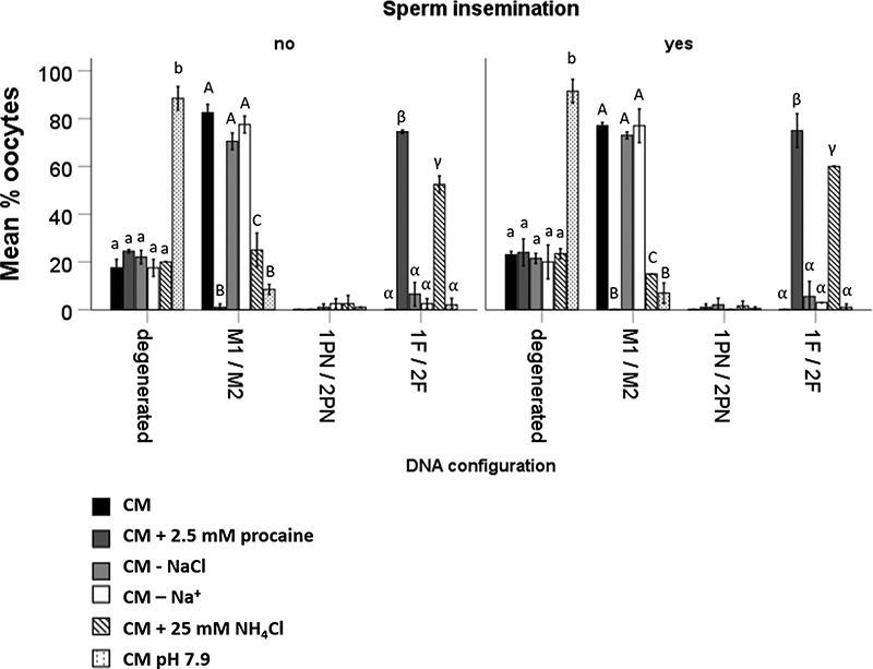
Figure 7.

Percentages of PD that showed (1) degeneration, (2) meiosis I stage (MI) or meiosis II stage (MII), (3) 1 PN or 2PN, and (4) 1 DNA fragment (1F) or 2 DNA fragments (2F) after 18 h incubation in CM, CM + 2.5 mM procaine, CM—NaCl, CM—Na+, CM + 25 mM NH4Cl, and CM pH 7.9. In general, oocytes exposed to procaine or NH4Cl rarely formed pronuclei, but instead exhibited condensed DNA fragments. Moreover, high medium pH exerted a degenerative effect on equine oocytes. Data represent mean (±SD) percentages of oocytes after incubation in CM, CM + 2.5 mM procaine, CM lacking NaCl, CM lacking Na+, CM + 25 mM NH4Cl, and CM at pH 7.9; n = 10 oocytes in each group, three replicates. Values that differ significantly (P < 0.05) within degenerated oocytes are indicated by small letters. Values that differ significantly between meiosis I and II oocytes are indicated by capital letters (P < 0.05). Values that differ significantly between oocytes containing DNA fragments are indicated by Greek letters (P < 0.05).
