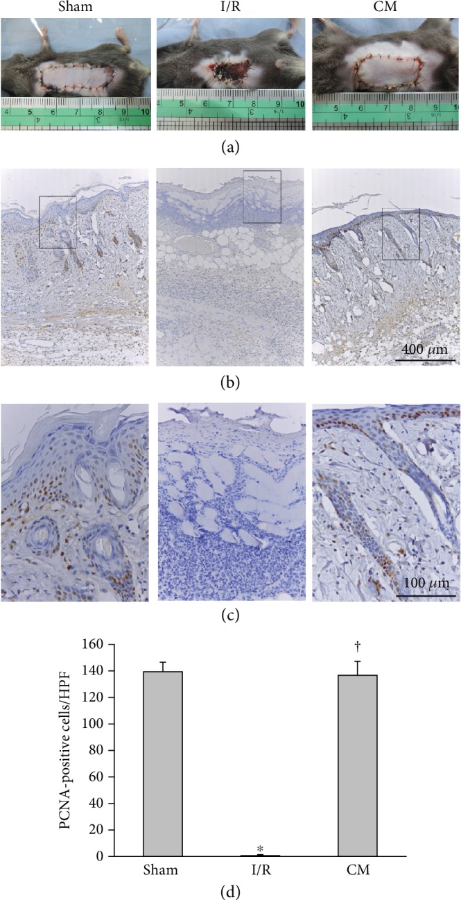
Figure 1.

ADSC-CM transplantation enhanced cell proliferation after I/R operation. (a) Flaps (4 × 1 cm2) of mice with ischemia induced by ligating long thoracic vessels for 3 h, which was then followed by blood reperfusion. ADSC-CM was administered into flaps through a subcutaneous route. Representative photographs of skin flaps on postoperative day 5 are shown. The necrotic areas of the I/R-induced skin flap were much larger than those of the sham group. In contrast, ADSC-CM (CM) treatment reduced the necrotic areas induced by I/R injury. (b) Immunostaining of PCNA. Bar = 400 μm. (c) Higher magnification of the area inside the rectangle in (b) is displayed. The PCNA-positive cells localized to the basal layer of the epidermis and the hair follicles. Bar = 100 μm. (d) Statistical analysis of PCNA-positive cells under high-power field (HPF) using ImagePro software. n = 6 for each group. ∗p < 0.05 versus the sham group; †p < 0.05 versus the I/R group.
