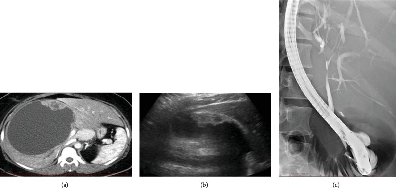
Figure 4.
Imaging findings in a patient with biliary mucinous cystic neoplasm. (a) CT image of a large BMCN in the right liver lobe with a hypodense solid component. (b) Ultrasound image of the same BMCN with a hypoechoic solid component by the wall. (c) ERCP image of the same patient showing s smooth stricture of the hepatic duct caused by extrinsic compression of the cyst.

