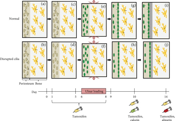
Figure 4.
Proposed effect of Prx1-driven primary cilium disruption on load-induced bone formation. Prx1-expressing periosteal progenitor cells (tan) and osteocytes (yellow) contain functional primary cilia (black) prior to experimentation (a, b). Tamoxifen is injected prior to loading, and periosteal progenitor primary cilia are uniquely disrupted in mutant mice (c, d). Ulnar loading is applied and both periosteal progenitors and osteocytes sense the mechanical stimulus. Periosteal progenitors are activated (green) by loading, regardless of whether their primary cilia are functional (e) or disrupted (f). The majority of activated progenitors migrate to the cortical surface (g, h). A continuous layer of matrix is deposited on the periosteal surface, but periosteal progenitors with disrupted cilia (j) produce less newly formed bone than those with functional cilia (i).

