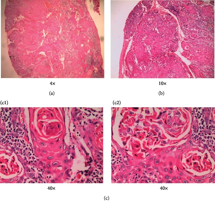
Figure 9.
(a) 4x and (b) 10x: histopathologic view of the lesion showing islands and sheets of malignant squamous epithelial cells infiltrating into the connective tissue. Areas showing severely atrophic epithelium with few saw toothed rete pegs and basal cell degeneration are also seen. (c1, c2) 40x: higher magnification showing neoplastic cells with numerous keratin pearl formations, nuclear and cellular pleomorphisms, and mitotic figures. Few dilated blood vessels were also noted.

