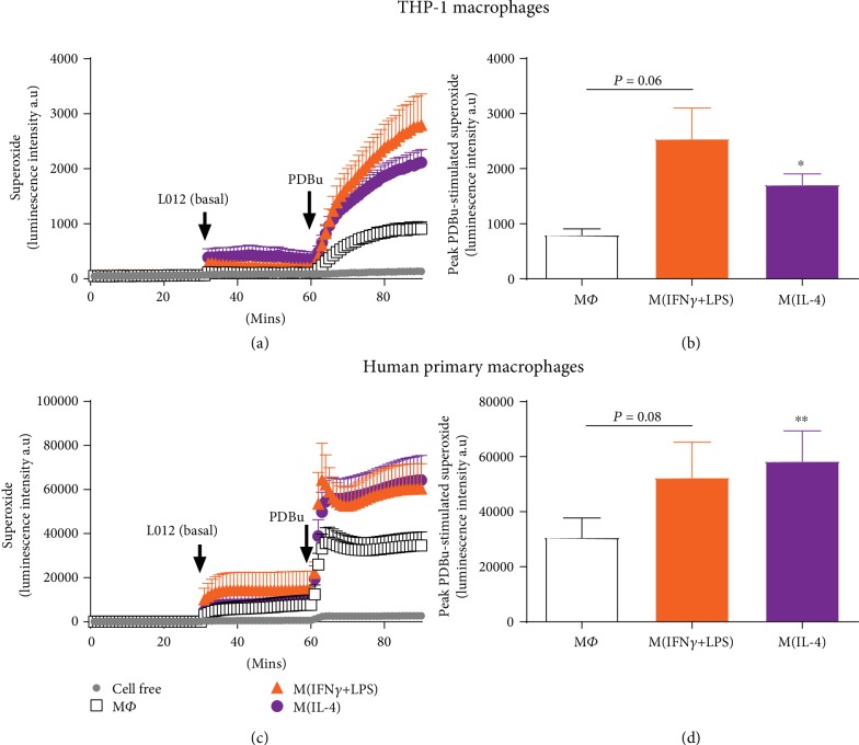
Figure 2.
PDBu-stimulated superoxide generation in activated human macrophages. PDBu-differentiated THP-1 macrophages (a, b) or M-CSF-differentiated human primary macrophages (c, d) were left untreated (MΦ) or treated with IFN-γ+LPS (M1) or IL-4 (M2) for 24 (THP-1) or 72 hours (primary macrophages), and superoxide generation was detected via L-012-enhanced chemiluminescence. Left hand side (LHS): average recordings demonstrating initial background readings (1-30 min), basal superoxide as detected following the addition of L-012 (100 μM; 31-60 min) and PDBu (10 μM)-stimulated superoxide generation (61-90 min) measured as luminescence intensity in arbitrary units (a.u) in (a) THP-1 and (c) primary macrophages. Right hand side (RHS): peak PDBu-stimulated (basal signal subtracted) superoxide generation in (b) THP-1 and (d) primary macrophages. All results presented as mean ± SEM, n = 7. ∗P < 0.05, ∗∗P < 0.01 vs. MΦ (1-way repeated measures ANOVA followed by Dunnett's post hoc test).

