Figure 1.
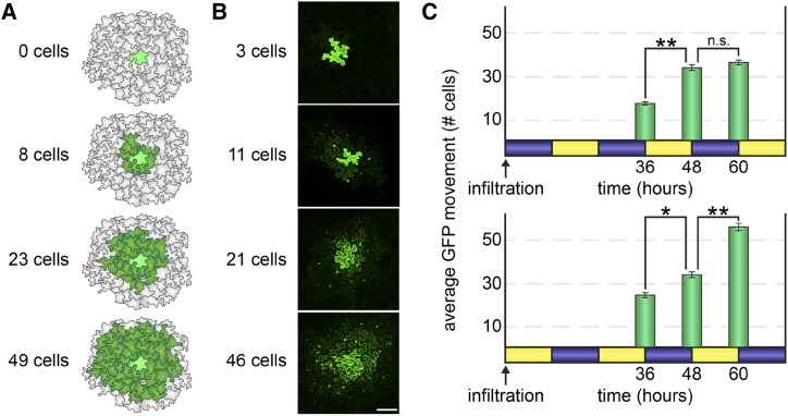
PD transport is higher during the day than at night. A, To measure rates of PD transport, we used a quantitative GFP movement assay. A very low inoculum of A. tumefaciens cells (less than 100 bacterial cells) was gently infiltrated by syringe into N. benthamiana leaves so that a handful of individual epidermal cells were transformed to express monomeric GFP. GFP spread to neighboring epidermal cells was then quantified 48 h after infiltration (or as indicated in each experiment). Illustrated examples of GFP movement are shown here. Movement is scored by counting the number of neighboring cells to which GFP has moved (dark green) from the transformed cell (bright green). B, Representative confocal microscopy images of the GFP movement assay. GFP fluorescence is brightest in the nuclei in cells neighboring the transformed cell. Bar = 100 μm. C, N. benthamiana leaves were agroinfiltrated to express GFP at either dusk (top) or dawn (bottom) in 5-week-old plants grown under 12-h-light (yellow)/12-h-dark (blue) cycles. GFP movement from the transformed cell was then assayed 36, 48, or 60 h later. In leaves infiltrated at dusk (top), GFP movement significantly increased during the second day but did not change during the third night. In leaves infiltrated at dawn (bottom), GFP movement somewhat increased during the second night but dramatically increased during the third day. *, P < 10−3 and **, P < 10−5; n.s., not significant.

