Figure 10.
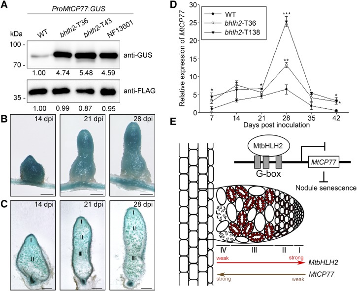
MtbHLH2 represses the expression of MtCP77. A, MtbHLH2 represses the expression of MtCP77 in M. truncatula. The 2-kb promoter region of MtCP77 was used for this assay. ProMtCP77:GUS, which carried an expression box of GFP-FLAG under the control of the CaMV35S promoter, was transformed into the wild type (WT) and bhlh2 mutants (bhlh2-T36, bhlh2-T43, and NF13601) by A. rhizogenes-mediated hairy root transformation. Composite transgenic plants were harvested, and proteins were extracted from ProMtCP77:GUS/wild type, ProMtCP77:GUS/bhlh2-T36, ProMtCP77:GUS/bhlh2-T43, and ProMtCP77:GUS/ NF13601 for immunoblot analysis. GFP-FLAG was used as a loading control. ImageJ software was used to quantify the immunoblot bands. B and C, GUS staining of MtCP77 expression in the nodules of ProMtCP77:GUS/bhlh2-T36. B, GUS staining of nodules on ProMtCP77:GUS/bhlh2-T36 hairy roots at 14, 21, and 28 dpi. C, Sections of nodules at 14, 21, and 28 dpi. The section thickness is 80 μm. The different zones of the nodules (I–III) are shown. Bars = 1 mm. D, The relative expression of MtCP77 in wild-type and bhlh2 mutant nodules (bhlh2-T36 and bhlh2-T138) at different time points after inoculation (7, 14, 21, 28, 35, and 42 dpi) with S. meliloti 1021 was quantified by RT-qPCR using MtACTIN as the reference gene. The error bars represent the ± sd of the means of two biological replicates, and asterisks indicate a significant difference between the wild-type and bhlh2 mutant nodules (one-way ANOVA followed by Tukey’s post-hoc test, *P < 0.05, **P < 0.01, ***P < 0.001). E, A proposed model for the regulation of MtbHLH2 during nodule senescence in M. truncatula. In the nodules, the expression of MtbHLH2 gradually decreases from the meristematic zone to the nitrogen fixation zone, whereas the expression of MtCP77 gradually increases. The binding of MtbHLH2 to the promoter of MtCP77 inhibits MtCP77 expression to delay nodule senescence. The different zones of the nodules (I–IV) are shown. I, Meristematic zone; II, infection zone; III, nitrogen fixation zone; IV, senescence zone.

