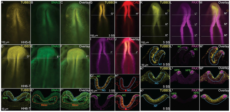
Fig. 2. TUBB3 is expressed in the developing neural plate prior to NC specification.

Immunohistochemistry (IHC) using antibodies against TUBB3 (yellow; A, C, D, F, D1, FI, G, G1, J, J1, K, K1–3, M, M1–3.), a neuron marker, SNAI2 (green; B, E, E1, F1), a definitive NC marker, N-cadherin (NCAD) (red; H, J, H1, J1), a cell adhesion molecule, and PAX7 (magenta, I, I1, J1, L, L1–3, M, M1–3), a neural crest progenitor marker. At stage HH5–6 (A–C) and stage 6–7 (D-F1) TUBB3 is expressed by cells in the neural plate (NP) and neural plate border (NPB) prior to the neural tube closure. Cranial mesenchyme/mesoderm is Meso. (G–J) IHC using antibodies against (G) TUBB3, (H) NCAD, (I) PAX7 and (J) Overlay in a 3 SS embryo. (G1-J1) Transverse sections through G-J. IHC for (K, K1–3) TUBB3, (L, L1–3) PAX7, and (M, M1–3) Overlay in a 5 SS embryo. TUBB3 is expressed in the NT (cranial), neural groove (NG, vagal), and NP at these axial levels. (A-F, G-J, K-M) are whole mounts with anterior to the top and posterior to the bottom while (D1-F1, G1-J1, K1-M3) are transverse sections with dorsal to the top and ventral to the bottom. These data suggest that TUBB3 protein may have early roles in addition to later fate determination. Scale bars are as marked. (For interpretation of the references to colour in this figure legend, the reader is referred to the Web version of this article.)
