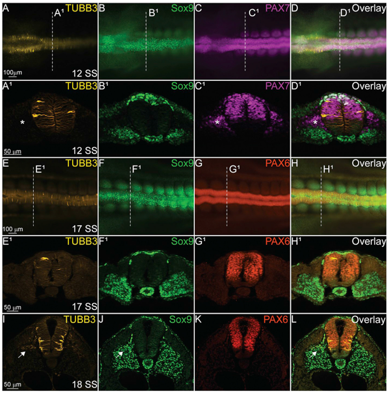
Fig. 5. TUBB3 does not co-localize with trunk NC cells.

IHC using antibodies against TUBB3 (yellow, A, A1, D, D1, E, E1, H, H1, I, L), SOX9 (green, B, B1, D, D1, F, F1, H, H1, J, L), PAX7 (magenta, C, C1, D, D1), and PAX6 (red, G, G1, H, H1, K, L) in the spinal cord of various staged embryos. TUBB3 is expressed intermittently in the developing spinal cord (A1, E1) at 12 SS and 17 SS, but expression becomes more stereotypic of differentiating spinal neurons at 18 SS (I). (A-D, E-H) Whole mount embryos with anterior to the left and posterior to the right. Dorsal side is facing reader. (A1-D1, E1-H1, I-L) Transverse sections with dorsal to the top and ventral to the bottom. Asterisks indicate TUBB3-/PAX7 + cells and arrows indicate TUBB3+/SOX9+ cells. Scale bars are as marked. (For interpretation of the references to colour in this figure legend, the reader is referred to the Web version of this article.)
