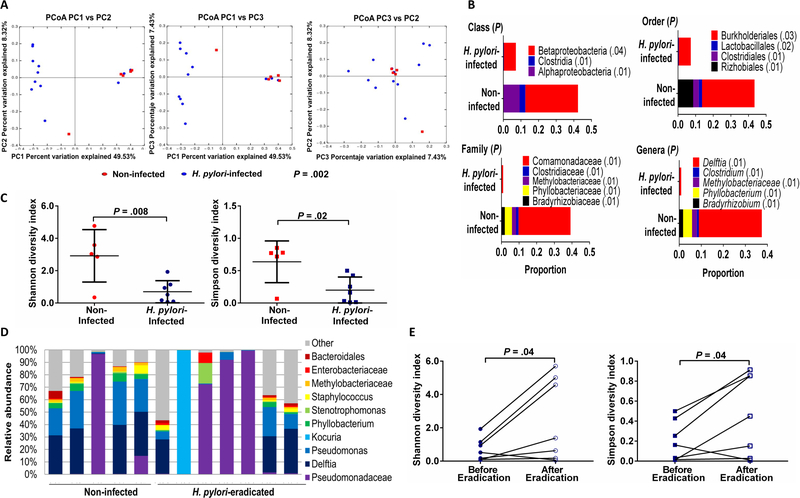
Figure 1.
Gastric microbiota in Venezuelan children is modified by H. pylori infection and reverts to that of non-infected children after H. pylori eradication. (A) β-diversity of the gastric microbiota in H. pylori-infected (n=11) and non-infected children (n=5) determined by permutational multivariate analysis of variance (PERMANOVA) and illustrated by unweighted UniFrac distances with each dot representing one subject in the principal coordinate analysis (PCoA) presented in 2-dimensional plots. P value determined by PERMANOVA test in QIIME. (B) Taxa abundances (when >1% of total bacterial DNA) at the class, order, family and genera levels among H. pylori-infected and non-infected children. P values determined by Kruskal-Wallis test with multiple comparison correction and false discovery rate analysis. When a genus could not be assigned, family is listed. (C) α-diversity of the gastric microbiota in H. pylori-infected (n=11) and non-infected children (n=5) using Shannon and Simpson indices. P values determined by Student’s t-test. (D) Taxa abundances of the 10 most abundant gastric bacteria (when >1% of total bacterial DNA) in children without H. pylori (5) and children in whom H. pylori was eradicated (n=7). P>.05, Kruskal-Wallis test. (E) α-diversity of the microbiota in H. pylori-infected children before (n=11) and after successful H. pylori eradication (n=7) using Shannon and Simpson indices. P values determined by Student’s t-test.

