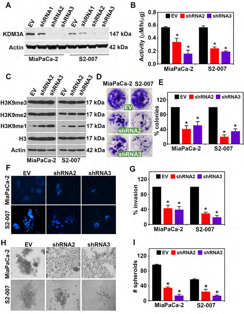
Figure 2. KDM3A knockdown inhibits malignant properties of PDAC cells.
(A) Western blot analysis for KDM3A in MiaPaCa-2 and S2–007 cells showing shRNA 2 and shRNA 3 efficiently lowered expression of KDM3A protein levels in comparison to an empty vector.
(B) Knockdown of KDM3A (shRNA 2 and shRNA 3) resulted in reduced KDM3A enzyme activity seen by measuring formaldehyde levels and increased substrate H3K9me1, a substrate for KDM3A, without altering H3, H3K9me2 and H3K9me3 levels in MiaPaCa-2 and S2–007 cells (p < 0.05).
(C) Knockdown of KDM3A (shRNA 2 and 3) reduced the specific substrate of KDM3A in particular H3K9me1, not the other substrate H3K9me2. H3K9me3 is not the specific substrate of KDM3A. Knockdown of KDM3A also inhibited (D-E) colony formation, (F-G) invasion and (H-I) spheroid formation in MiaPaCa-2 and S2–007 cells in PDAC cell lines

