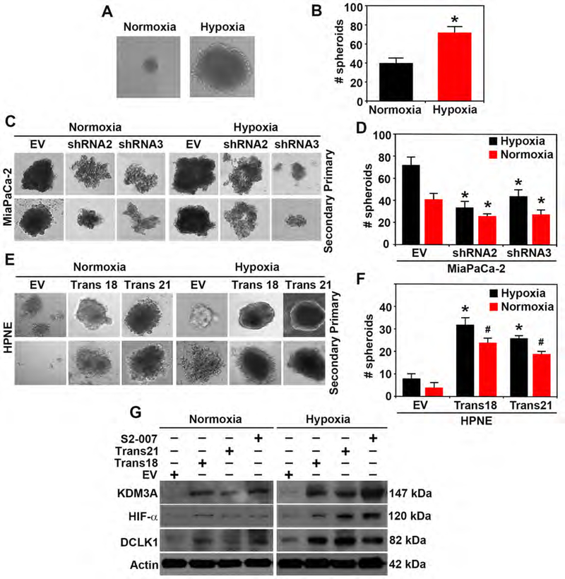
Figure 4. Hypoxia induces pancosphere formation and increased KDM3A in PDAC and transformed HPNE cells.
(A & B) Pancosphere formation (size and number) is significantly increased during hypoxic conditions (p < 0.05).
(C-D) Primary and secondary spheroids formation (size and number) are significantly inhibited by silencing of KDM3A (shRNA2 and shRNA3) in hypoxic and normoxic conditions in MiaPaCa2 PDAC cells (p < 0.05).
(E-F) Overexpression of KDM3A in HPNE non-cancerous cell (Trans18 and Trans21 transformed clones) showed a significant increase in size and number of spheroids (p < 0.05) in both normoxic and hypoxic conditions in comparison to empty vectors (EV).
(G) Western blot analysis of KDM3A, HIF-α and DCLK1 in S2–007, Trans18 and Trans21 HPNE in comparison to EV during normoxic and hypoxic condition demonstrating hypoxia increased the expressions of those proteins.

