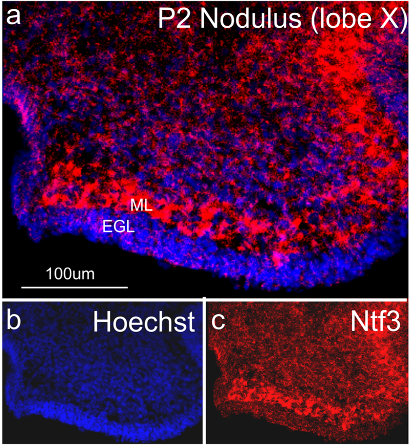
Fig. 6.
Ntf3 expression in a thick sagittal section of the cerebellum. The RNAscope technique developed here works equally well on thick sections to show the expression of the neurotrophin Ntf3 in the molecular layer (ML) of the cerebellum. The proliferating granule cell precursors in the external granule layer (EGL) show very limited expression compared to the cells in the molecular layer (ML) that at this stage includes Purkinje cells

