
Figure 2.
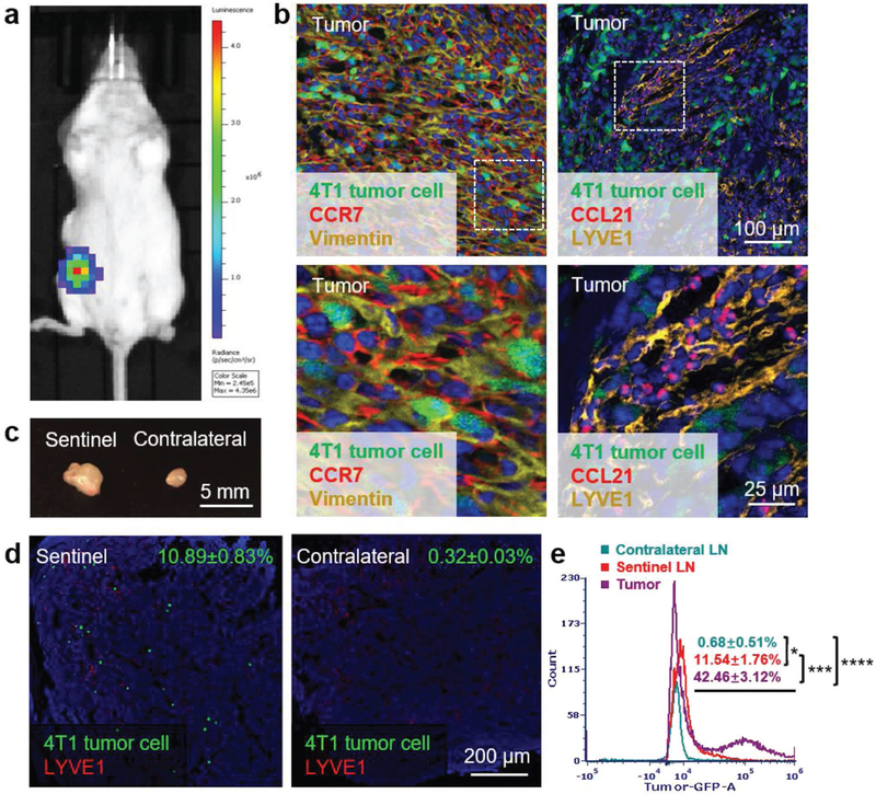
Murine 4T1 breast cancer is a highly metastatic orthotopic TNBC model with CCR7 overexpression. a) Bioluminescence imaging of 4T1 breast cancer orthotopic TNBC model. b) Immunofluorescence staining of CCR7 and vimentin expressed in tumor (left panel, green: 4T1 tumor cells, red: CCR7, yellow: vimentin, blue: nucleus); immunofluorescence staining of lymphatic vessel marker LYVE1 and cytokine CCL21 in tumor (right panel, green: 4T1 tumor cells, red: CCL21, yellow: LYVE1, blue: nucleus). c) Volume comparison of sentinel and contralateral LNs on day 10 after tumor inoculation. d) Immunofluorescence staining of 4T1 tumor cell metastasis in sentinel and contralateral LNs (green: 4T1 tumor cells, red: LYVE1, blue: nucleus). e) Flow cytometry measuring green fluorescence of 4T1 cells in tumor, sentinel and contralateral LNs (*p < 0.05; ***, p < 0.001; ****, p < 0.0001; n=6).