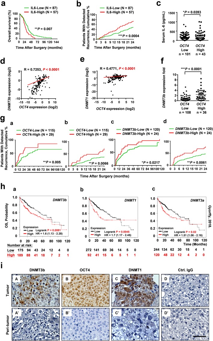
Fig. 1.
Correlation between serum IL-6 and tissue DNMT3b/OCT4 with the patient prognosis of human HCC. The overall survival (OS) (a) and early tumor recurrence (within 24 months) (b) of patients after HCC resection based on high or low serum IL-6 level by Kaplan-Meier analysis (n = 144, cutoff for high IL-6 concentration was 150 pg/mL). After normalization with the corresponding peritumor (PT) tissue sample, the expression levels (either high [T/PT ≧ 2 or low [T/PT < 2]) of OCT4 were assessed. c The differences in serum levels of IL-6 between HCC patients with low OCT4 expression (T/PT < 2-fold; n = 101) and high OCT4 expression (T/PT ≥ 2-fold; n = 43) are shown. Positive correlations by Spearman analysis between expression levels of OCT4 with DNMT3b (R = 0.7253) (d) and DNMT1 (R = 0.4771) (e) in HCC tissues. n = 144. The differences in DNMT3b between HCC patients with low OCT4 expression (T/PT < 2-fold; n = 108) and high OCT4 expression (T/PT ≥ 2-fold; n = 36) (f) Statistical significance was assessed by the Mann–Whitney U test. (*P < 0.05; ***P < 0.001). g The Kaplan-Meier curves of tumor recurrence (120 months) or early recurrence (24 months) in relation to the transcriptional levels of OCT4 (n = 144), DNMT3b (n = 144) in human HCC tissue (*P < 0.05, **P < 0.01). h The OS analysis of DNMT expression in HCC using The Cancer Genome Atlas (TCGA) dataset by Kaplan-Meier analysis (n = 364). The top tertile was defined as the high DNMTs expression cohort and the remaining patients were defined as the low DNMTs expression cohort. i The expression and localization of DNMT3b, OCT4, and DNMT1 in HCC tissues by immuno-histochemical staining. (Bar, 100 μm)

