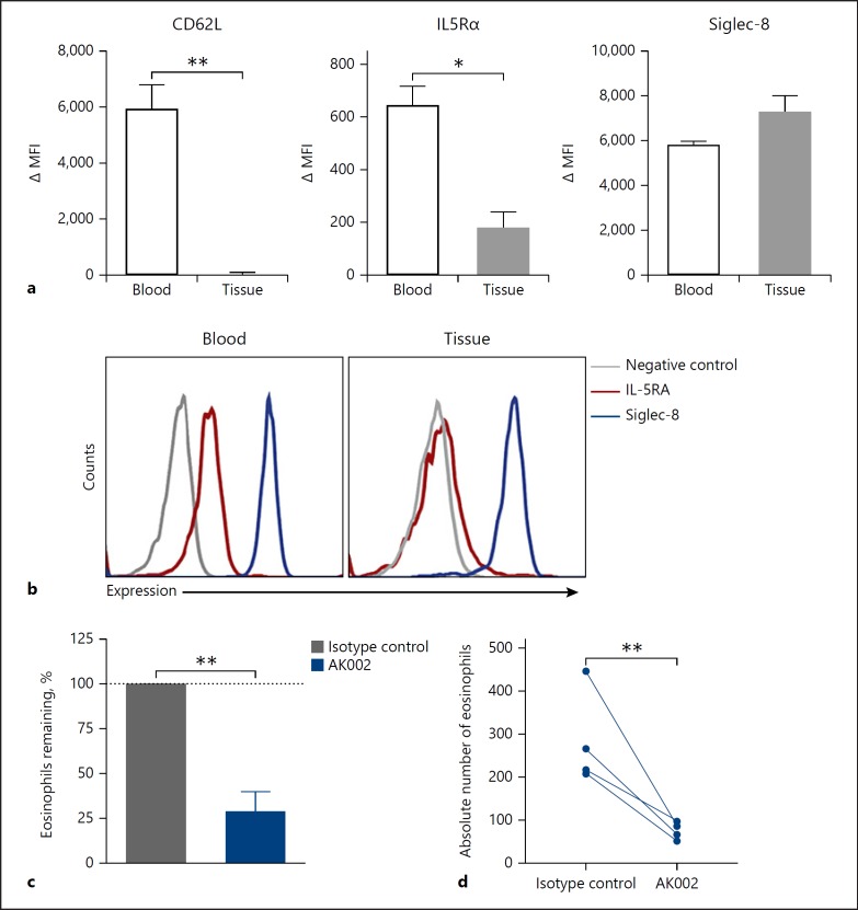
Fig. 5.
AK002 reduces human tissue eosinophils ex vivo. a Surface expression of CD62L, IL-5Rα and Siglec-8 plotted as ΔMFI on blood (black) and lung tissue (gray) eosinophils (mean ± SD of 4 donors). b Representative histograms of surface expression of IL-5Rα (red), Siglec-8 (blue) or fluorescence minus one negative control (gray) on blood and lung tissue eosinophils. c, d Dissociated human tissue was incubated overnight with 1 μg/mL isotype control (gray) or AK002 (blue). Eosinophils were counted by flow cytometry and plotted as the (c) percent eosinophils remaining or as (d) absolute eosinophil counts. The percent eosinophils remaining was calculated by normalizing to the percent of CD45+ eosinophils in the isotype control-treated wells to 100% (mean ± SD of 4 donors). * p < 0.05; ** p < 0.01. MFI, median fluorescence intensity; IL5Rα, interleukin-5 receptor α; Siglec, sialic acid-binding immunoglobulin-like lectin.

