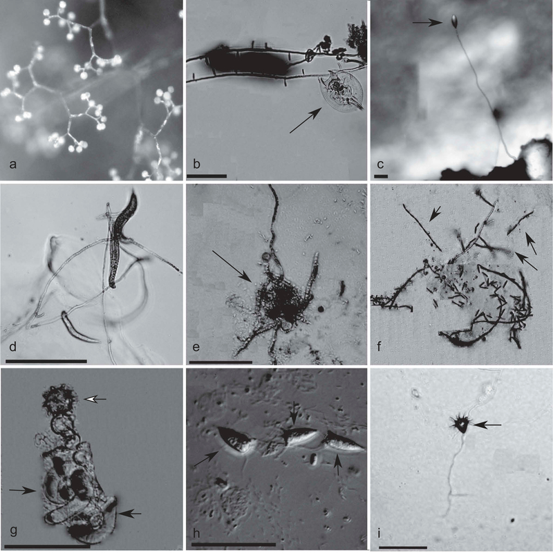
Figure 2.
A. Sporangiophore of Piptocephalis sp. Photograph taken at 80× with a stereomicroscope. B. Rotifer (black arrow) trapped on hyphae of Zoophagus insidians. Photograph taken at 100×. C. Conidiophore and conidium (black arrow) of Stylopage hadra. Photograph taken at 80× with a stereomicroscope. D. Hyphae of Zoophagus pectospora with trapped Bunoema nematodes. Photograph taken at 100×. E. Amoeba (black arrow) invaded and partially digested by the Ohio isolate of Zoopage sp. Photograph taken at 400×. F. Catenulated conidia chains (black arrows) of the Arizona Zoopage sp. isolate. Photograph taken at 100× through the agar plate. G. Thallus (black arrows) and zygospore (white arrow) of Cochlonema odontosperma. Photograph taken at 200×. H. Conidia (black arrows) of Acaulopage tetraceros. Photograph taken at 400× with DIC. I. Conidium (black arrow) of Acaulopage acanthospora. Photograph taken at 100× through the agar plate. Bars = 50 μm.

