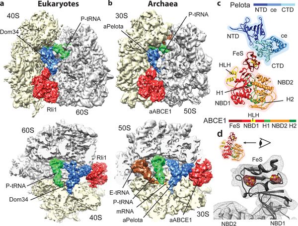
Fig. 1: The ribosome-bound Pelota-ABCE1 complex.
(a, b) Cryo-EM reconstructions of the eukaryotic SL-RNC-Dom34-Rli1 and the archaeal 70S-aPelota-aABCE1 complex at 7.2 Å and 6.6 Å resolution, respectively. Extra densities were observed for Dom34/aPelota and Rli1/aABCE1 in the canonical factor binding site as well as for P-site tRNA, E-site tRNA and mRNA. The upper section represents side views, the lower section top views, where large and small subunits were cut. (c) Homology model for ribosome-bound Pelota and ABCE1 in transparent density. The individual domains are color-coded as in the schematic representation of domain organization. N-terminal Domain (NTD), central domain (ce) and C-terminal domain (CTD) are indicated; FeS indicates iron-sulfur cluster domain; NBD1 and 2 indicate nucleotide binding domain 1 and 2; HLH indicates helix-loop-helix motif; H1 and H2 indicates hinge 1 and hinge 2 domain. (d) Zoom on the FeS domain of aABCE1. The density for the two [4Fe-4S]2+ is displayed in red mesh at high contour level.

