Fig. 4:
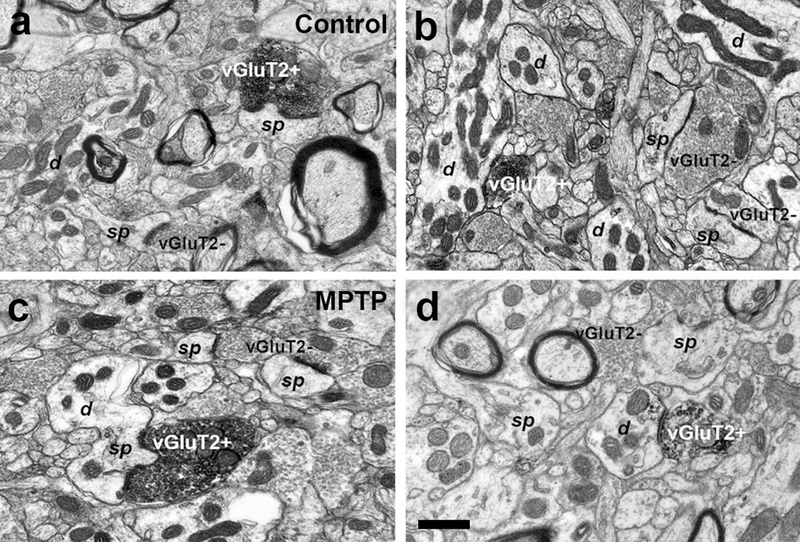
Electron micrographs of vGluT2-positive (vGluT2+) terminals forming asymmetric synapses with dendrites (d) and dendritic spines (sp) in the caudate (b, c) and putamen (a, d) of control (a, b) and MPTP-treated-parkinsonian monkeys (c, d). In the same field, vGluT2- negative (vGluT2-) terminals form asymmetric synapses with dendritic spines. Scale bar in a (applies to b and c) and in d = 0.5μm

