
Fig. 6 :
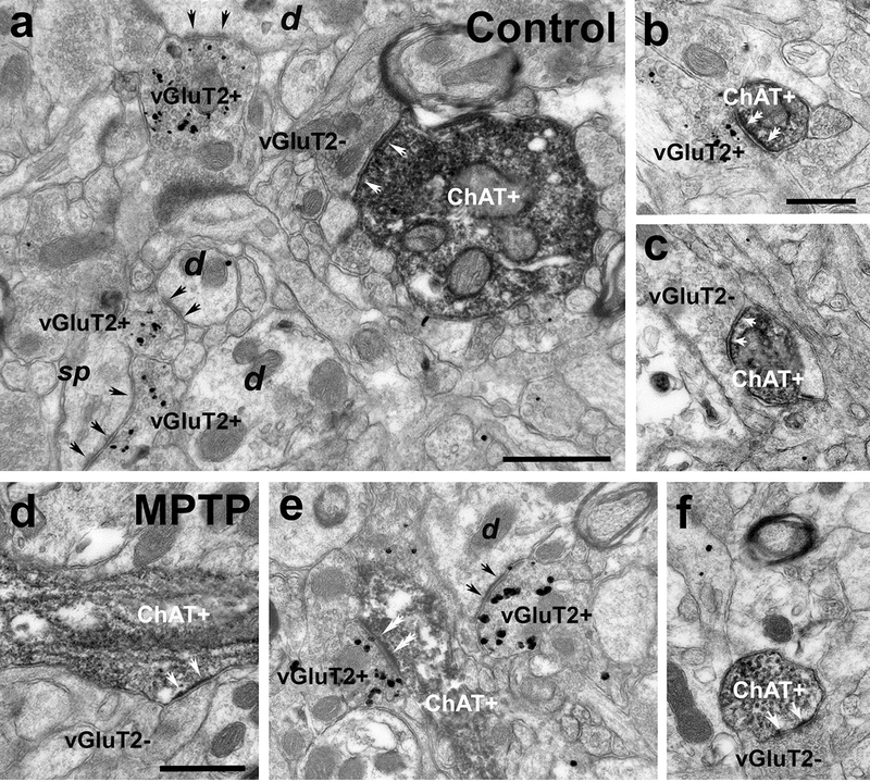
Electron micrographs of ChAT-immunostained (peroxidase) dendrites and vGluT2-immunolabeled (silver-intensified gold particles) terminals in the putamen (a, b, d, e) and caudate (c, f) of control (a, b, c) and MPTP-treated monkeys (d, e, f). (a) A large-sized ChAT+ dendrite (diameter >1μm) forms an asymmetric synapse (white arrows) with a vGluT2-negative (vGluT2-) terminal. In the same field, some vGluT2-positive (vGluT2+) terminals form asymmetric synapses (black arrows) with non-labeled dendrites (d) and a dendritic spine (sp). (b) A vGluT2+ terminal forms an asymmetric synapse with a small-sized (diameter <0.5μm) ChAT+ dendrite (double white arrows). (c) Medium-sized (diameter between 0.5–1μm) ChAT-immunostained dendrite receiving an asymmetric synapse from a vGluT2-negative (white arrows). (d-f) Large- (d) and medium-sized (e, f) ChAT+ dendrites form asymmetric synapses with vGluT2- (white arrows in d and f) and vGluT2+ (double white arrows in e) terminals. Scale bars in a = 0.5μm, in b (applies to c) = 0.5 μm and in d = 0.5μm (applies to d and f)