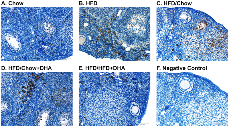
Figure 5. Ovarian Macrophage Infiltration.
Macrophages present in the ovary were identified using the CD68 marker in Chow (N = 5), HFD (N = 5), HFD/Chow (N = 5), HFD/Chow+DHA (N = 4), and HFD/HFD+DHA (N = 5) mice. Representative images are shown for (A) Chow, (B) HFD, (C) HFD/Chow, (D) HFD/Chow+DHA, (E), HFD/HFD+DHA, and (F) negative no-primary antibody control.

