Figure 1.

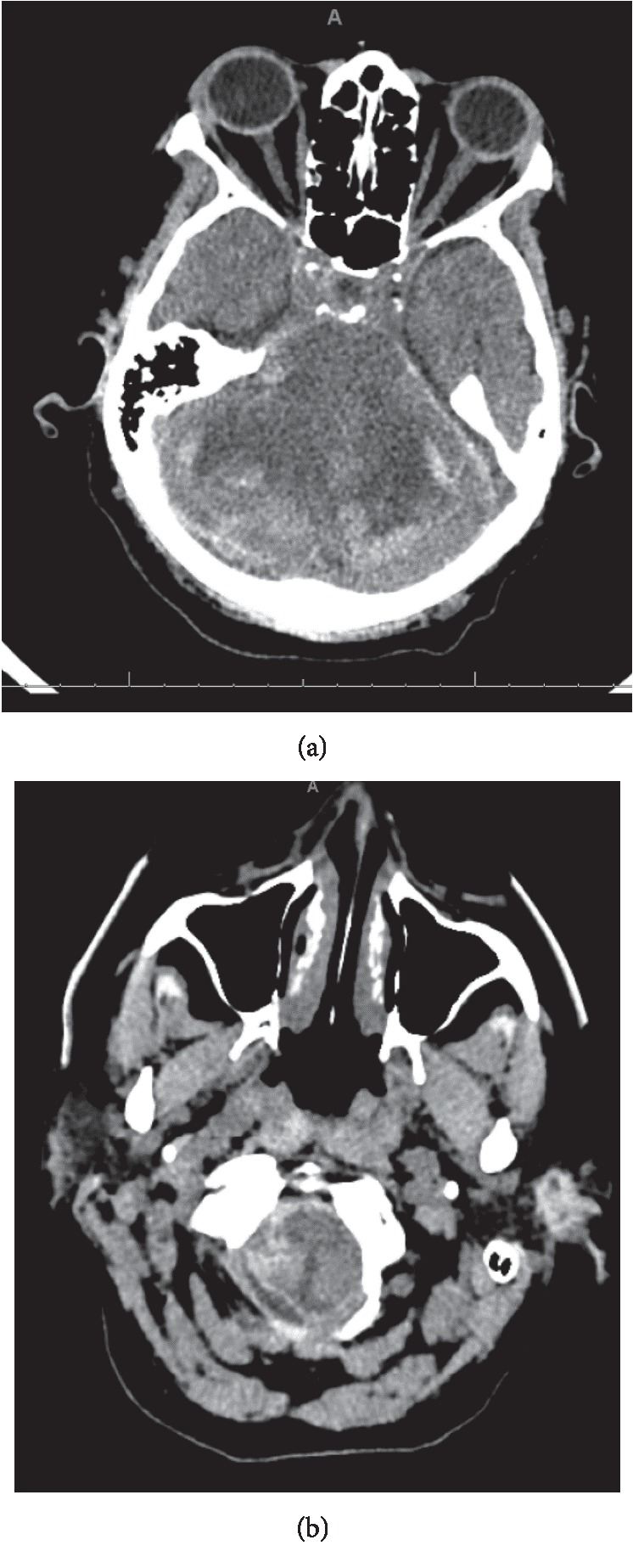
Peripheral hemorrhage in the cerebellar folia with associated areas of hypodensity (a), along with hypodensity and hemorrhage within the brainstem (b), both consistent with ischemia.

Peripheral hemorrhage in the cerebellar folia with associated areas of hypodensity (a), along with hypodensity and hemorrhage within the brainstem (b), both consistent with ischemia.