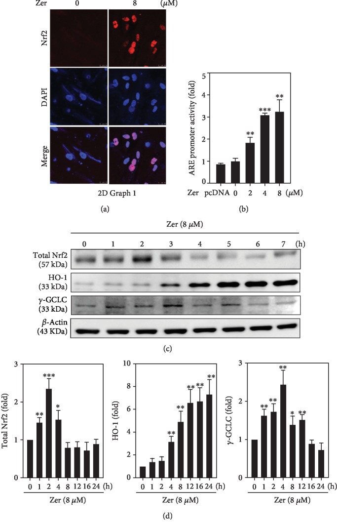
Figure 5.
Effect of ZER on ARE promoter activation and subsequent expression of HO-1 and γ-GCLC proteins in HSF cells. (a) Cells were treated with ZER (8 μM for 2 h) and subcellular localization of Nrf2 was determined using immunostaining method. (b) HSF cells were cotransfected with pGL3-ARE and treated with various concentrations of ZER (2-8 μM for 2 h) to measure the percentage of ARE promoter activity. Data was presented as fold over increase in the percentage of ARE promoter activity. (c, d) The effect of ZER treatment (8 μM) on the expression of total Nrf2 and antioxidant proteins (HO-1 and γ-GCLC) at different time points (0, 1, 2, 4, 8, 12, 16, or 24 h) was measured using western blot method against β-actin as an internal control. Results from three or more experiments were presented as mean ± SD, and the statistical significance was considered as ∗p < 0.05, ∗∗p < 0.01, ∗∗∗p < 0.001 compared to untreated control cells.

