Figure 5.
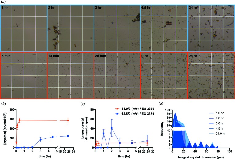
Observing a 100 µl FutA batch crystallization over 24 h. (a) The growth of two FutA batch crystallization experiments, the top (blue) in 0.2 M NaSCN, 12.5%(w/v) PEG 3350 and the bottom (red) in 0.1 M Tris pH 7.1, 38.0%(w/v) PEG 3350. The pictures show aliquots viewed in a hemocytometer. The white boxes in the images have dimensions of 250 × 250 µm. (b), (c) Demonstrations of how the mean number of crystals and longest dimension change over time. (d) A histogram of crystal size over 24 h for the 12.5%(w/v) PEG 3350 condition.

