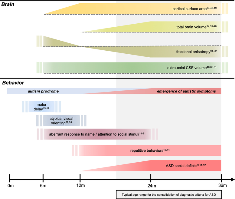
Figure 1. Summary of neuroimaging findings in ASD in the context of emerging behaviors from infancy through toddlerhood.
Brain changes in ASD precede the development of the defining diagnostic features of the disorder, and are temporally associated with behavioral changes in the first year of life that are both specific and non-specific to ASD. Aberrant white matter integrity (fractional anisotropy) and increased extra-axial cerebrospinal fluid (CSF) volumes are detectable as early as 6 months of age in infants who go on to develop ASD, concurrent with motor and sensory delays. Surface area hyper-expansion in the first year of life precedes brain overgrowth in the second year, during which time ASD symptoms become apparent and begin to consolidate, while brain phenotypes remain relatively stable. Findings presented in the figure are those which are supported by multiple study paradigms (reference numbers noted in the figure), including at least one longitudinal study per phenotype. Double bars indicate that the start and/or end point of the trajectory is unknown or not well documented in the literature. Dashed lines in the top panel represent a reference to typical brain development, where bars above or below the dotted line indicate the brain phenotype is either increased or decreased relative to controls, respectively. For example, fractional anisotropy in ASD is increased at 6 months, not significantly different at 12 months, and decreased from 24 to 36 months when compared to controls. Repetitive behaviors and ASD social deficits are shown to continue past 36 months without citations, as these are diagnostic features that are, by definition, present in individuals with an ASD diagnosis.

