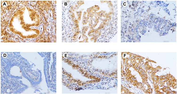
Figure 3.
Immunohistochemical staining of PHD2 in endometrial tissues. (A) High expression of PHD2 in normal endometrium (×400); (B) moderate expression of PHD2 in atypical endometrial hyperplasia (×400); (C) low expression of PHD2 in endometrial cancer (×400); (D) low expression of HIF-1α in normal endometrium (×400); (E) moderate expression of HIF-1α in atypical endometrial hyperplasia (×400); (F) high expression of HIF-1α in endometrial cancer (×400).

