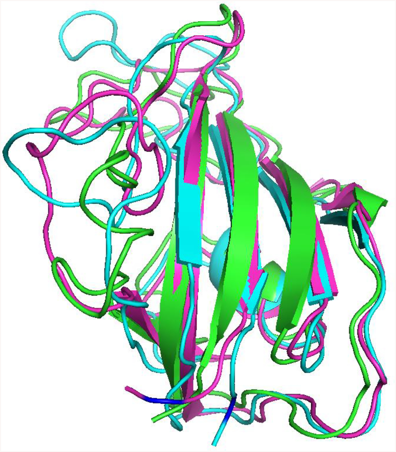
Figure 5. Molecular dynamics analysis of the rapamycin-induced FKBP-25 F145A/I223P double mutant disorder-to-order transition.
Wild type, ligand-free, and ligand-bound mutant proteins are shown in green, cyan, and magenta, respectively. The I223P mutation is indicated in blue. The overall protein structure is preserved between all three proteins. The β-sheet secondary structure in the region of the mutation is lost in the non-ligand-bound mutant protein and is recovered in the ligand-bound form of the mutant protein.

