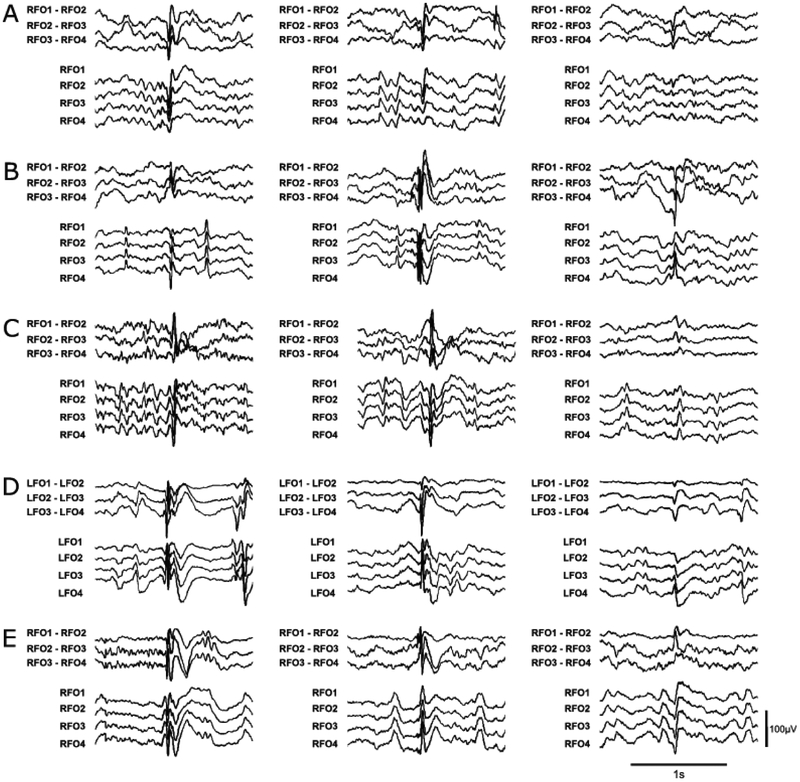
Figure 1:
Representative examples of “definite” and “indeterminate” IEDs for 5 patients, including (A) Patient #4; (B) Patient #9; (C) Patient #25; (D) Patient #26; and (E) Patient #28. The left and middle columns show “definite” IEDs, while the right column shows “indeterminate” IEDs. Amplitude and time scales for all examples are shown in the bottom right corner. Patient numbers correspond to those shown in Supplementary Table 1.

